Screen 1 of 12
Welcome back
Screen 1
Three kinds of blood vessel
- Blood flows round your body in tubes called blood vessels.
- Arteries carry blood away from the heart. Veins carry it back to the heart.
- Tiny capillaries link them, close to almost every cell.
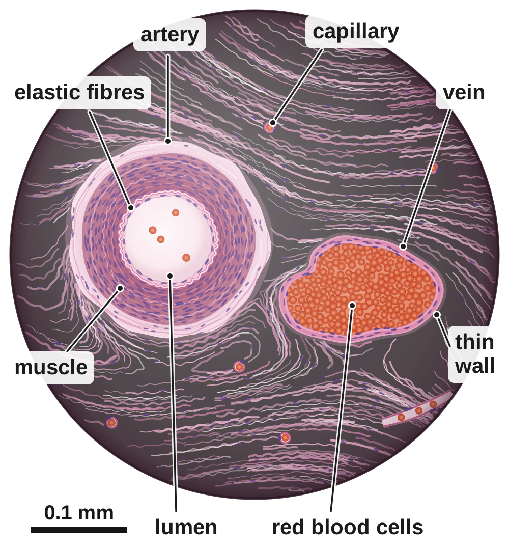
Where the slice comes from. Red = oxygenated. Blue = low in oxygen.
Screen 2
A real slice and a diagram
- A real slice is stained, and the vein's thin wall is often squashed.
- A diagram is simplified on purpose: neat circles, flat colours and the capillary drawn bigger.
- Real blood is never blue; veins look blue through skin only.
Tap a difference to see it on both pictures.
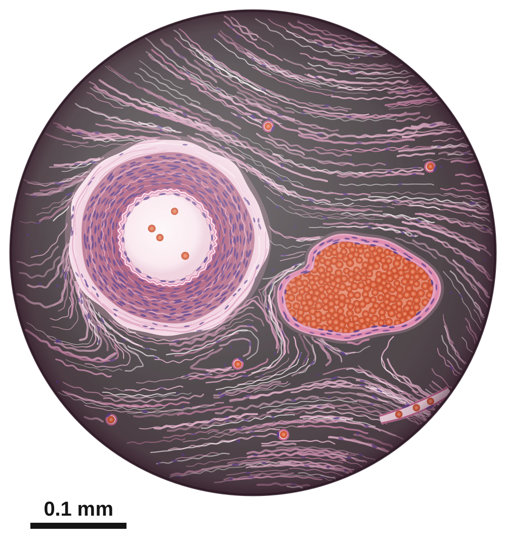
A real slice
left: artery · right: vein
A diagram
left: artery · right: vein
Real blood is never blue; veins look blue through skin only.
Screen 3
Built for the job
- Artery: a thick wall of muscle and elastic fibres, and a narrow lumen.
- Vein: a thin wall, a wide lumen and valves.
- Capillary: a wall just one cell thick.
Tap a part to see its job.
Screen 4
Arteries: stretch and recoil
- The heart pumps blood into the arteries at high pressure, in surges.
- Elastic fibres stretch as each surge passes, then recoil. You feel this as your pulse.
- A thick wall of muscle makes the artery strong.
Red = oxygenated. Blue = low in oxygen. Real blood is never blue.
Screen 5
Checkpoint 1 of 3
Quick check on screens 1 to 4. Tap an answer.
1.In a slice seen under a microscope, how can you tell the artery from the vein?
2.What is the lumen of a blood vessel?
3.What do the elastic fibres in an artery wall do?
Screen 6
Veins and valves
- Blood in veins is at low pressure, so veins have a thin wall and a wide lumen.
- Valves stop blood flowing backwards.
- When your leg muscles contract, they squeeze the veins and push blood towards the heart.
Each squeeze pushes blood up through the open valve. The valve below shuts, so the blood cannot go back down.
Blue = low in oxygen. Real blood is never blue; veins look blue through skin only.
Screen 7
Capillaries: exchange with cells
- Capillary walls are one cell thick, so substances diffuse only a short distance.
- Oxygen and glucose diffuse into the body cells. Carbon dioxide diffuses into the blood.
- Red blood cells squeeze through in single file, close to the cells.
The wall is one cell thick, so every substance has only a short distance to diffuse.
Screen 8
Checkpoint 2 of 3
Quick check on screens 6 and 7. Tap an answer.
1.Which type of blood vessel has valves along its length?
2.Why do veins have a thin wall and a wide lumen?
3.What is the wall of a capillary made of?
Screen 9
Pressure from artery to vein
- Blood pressure is highest in the arteries, and rises and falls with each heartbeat.
- It falls as blood passes through the capillaries. In the veins it is low and steady.
| Feature | Artery | Vein | Capillary |
|---|---|---|---|
| Carries blood | away from the heart | back to the heart | from arteries to veins |
| Pressure | high, with a pulse | low and steady | falling |
| Wall | thick: muscle and elastic fibres | thin | one cell thick |
| Lumen | narrow | wide | very narrow |
| Valves | no | yes | no |
Read it: in the arteries the pressure peaks at about 16 kPa with each heartbeat.
Tap a type of blood vessel, or follow the blood.
Screen 10
Artery, vein or capillary?
- Use the clues: the wall, the lumen, the valves and the direction the blood flows.
- Name a vessel by its direction, not its oxygen: the pulmonary artery carries blood low in oxygen.
Score: 0 out of 6
Red = oxygenated. Blue = low in oxygen. Real blood is never blue.
Thick wall. Narrow lumen. Blood surges through at high pressure.
Thin wall. Valves along its length.
Wall one cell thick. Red blood cells pass in single file.
Carries blood low in oxygen from the heart to the lungs.
Carries oxygenated blood from the lungs to the heart.
Lets oxygen and glucose diffuse out to muscle cells.
Screen 11
Rate of blood flow
- Rate of blood flow = volume of blood ÷ time, in cm³ per minute.
- Change litres to cm³ (× 1000) and seconds to minutes (÷ 60) first.
- It is a volume per minute, not the heart rate.
Example: 360 cm³ in 2 minutes. 360 ÷ 2 = 180 cm³ per minute.
A model: the meter adds up the volume of blood that passes in a measured time.
Screen 12
Checkpoint 3 of 3
Quick check on screens 9 to 11. Tap an answer.
1.In which blood vessels is the blood pressure highest?
2.The pulmonary artery carries blood low in oxygen. Why is it still called an artery?
3.200 cm³ of blood flows through an artery in 4 minutes. What is the rate of blood flow?
Quiz
Quiz: B2 L7 Blood vessels
10 questions. Tap an answer. Your first answer counts towards your score.
In a real stained slice, the vein often looks squashed and uneven. Why?
Which of these describes an artery?
Why does an artery have a thick layer of muscle in its wall?
You can feel a pulse at your wrist. What causes it?
How do the leg muscles help blood get back to the heart?
Which substance diffuses from the body cells into the blood in a capillary?
Capillaries are so narrow that red blood cells pass through in single file. How does this help?
The blood pressure in an artery is about 16 kPa. In a vein near the heart it is about 1 kPa. What does this show?
Which statement about blood vessels is true?
0.7 litres of blood flows through an artery in 30 seconds. What is the rate of blood flow?
Exam questions
The same five questions as the B2 L7 Exam questions worksheet. 20 marks.
Write your answer, then open the mark scheme and tick each point you made.
01Figure 1 shows three blood vessels, A, B and C, cut across.
They are not drawn to the same scale.
01.1Complete Table 1.
Choose answers from the box.
| Blood vessel | Type of blood vessel |
|---|---|
| A | |
| B | |
| C |
01.2Which structure is found along veins but not along arteries?
Tick (✓) one box.
Mark scheme
- (1)
- (1)
- (1)
- (1)
01.1: 1 mark for each correct row. "nerve" and "valve" are not used. If more than one word is written in a box, do not give that mark.
Model answer01.1 A vein; B artery; C capillary. 01.2 Valves.
02Doctors measured the blood flow in the main artery in a person's leg.
They measured it at rest and during exercise.
Table 2 shows the results.
| Activity | Volume of blood in cm³ | Time in minutes | Rate of blood flow in cm³/min |
|---|---|---|---|
| At rest | 900 | 3 | X |
| During exercise | 2400 | 2 | 1200 |
02.1Calculate value X in Table 2.
Use the equation:
rate of blood flow = volume of blood ÷ time
02.2How many times greater is the rate of blood flow during exercise than at rest?
Use your answer to Question 02.1.
02.3Suggest why the rate of blood flow to the leg muscles is greater during exercise.
Mark scheme
- (1)
- (1)
- (1)
- (1)
- (1)
02.1: correct answer with no working = 2 marks.
02.2: allow 1200 ÷ their 02.1 answer, correctly calculated. allow "4 times".
02.3: allow "the muscles need more energy (released by respiration)" for the first mark. allow "more carbon dioxide needs to be removed" for the second mark. max 2. ignore "the heart beats faster" (that is how, not why). do not accept "more energy is made / produced / created" (that point scores 0, others can still score).
Model answer02.1 900 ÷ 3 = 300 cm³ per minute. 02.2 1200 ÷ 300 = 4 times. 02.3 The leg muscle cells respire faster during exercise, so they need more oxygen and glucose to be delivered by the blood.
03Figure 2 shows a vein in the leg, cut lengthways.
03.1Name structure X.
03.2Explain how structure X keeps blood flowing towards the heart.
Mark scheme
- (1)
- (1)
- (1)
03.1: allow "valves". do not accept "elastic fibres" / "muscle".
03.2: allow "it stops blood flowing backwards" for 1 mark if neither point is given in full. do not accept "the valve pumps blood (to the heart)".
Model answer03.1 A valve. 03.2 The valve opens to let blood flow up towards the heart. If the blood starts to flow back down, the valve shuts, so the blood cannot flow backwards.
04A student wrote:
'Arteries always carry oxygenated blood.'
04.1Name one artery that shows the student is wrong.
04.2Blood vessels are named by the direction the blood flows in them.
Give the direction of blood flow in an artery and in a vein.
Mark scheme
- (1)
- (1)
04.1: do not accept "pulmonary vein" / "aorta" / "coronary artery".
04.2: both needed for the mark. do not accept answers in terms of oxygen.
Model answer04.1 The pulmonary artery. 04.2 Arteries carry blood away from the heart; veins carry blood back to the heart.
05There are three types of blood vessel: arteries, veins and capillaries.
Explain how the structure of each type of blood vessel is related to its function.
Mark scheme
Level of response. Read your answer, choose the level that fits best, then pick your mark.
| Level | Marks | Descriptor |
|---|---|---|
| 3 | 5–6 | A detailed and coherent explanation covering all three types of blood vessel. Each has at least one structural feature clearly linked to its function, with logical links, for example: wall one cell thick → short diffusion distance → fast exchange with the cells. |
| 2 | 3–4 | Relevant features of at least two types of blood vessel are given, and some are linked to their function. The links may be incomplete or not fully explained. |
| 1 | 1–2 | Simple statements of structure or of function, for example "veins have valves" or "arteries carry blood away from the heart", with little or no link between them. |
| 0 | 0 | No relevant content. |
Indicative content
- arteries carry blood away from the heart at high pressure
- arteries have a thick wall / thick layer of muscle, so the wall is strong enough to withstand the high pressure (without bursting)
- arteries have elastic fibres, which stretch as each surge of blood passes and then recoil, pushing the blood on / keeping the pressure up
- arteries have a narrow lumen, which keeps the pressure high
- veins carry blood back to the heart at low pressure
- veins have a thin wall, because the pressure is low
- veins have a wide lumen, so blood flows easily at low pressure
- veins have valves, which stop blood flowing backwards / keep blood flowing towards the heart
- capillaries link arteries to veins and exchange substances with the body cells
- capillaries have a wall one cell thick, so the diffusion distance is short
- capillaries are very narrow, so blood passes close to the cells (red blood cells in single file)
- there are many capillaries / a large network, giving a large surface area for diffusion
- oxygen and glucose diffuse into the cells; carbon dioxide diffuses into the blood
- do not accept "capillaries have a cell wall", "arteries carry oxygenated blood" as the function of arteries, or "the muscle pumps the blood" (these statements do not score; the rest of the answer can still score)
Your mark:
Model answer (Level 3)Arteries carry blood away from the heart at high pressure. They have a thick wall of muscle, so they are strong enough to withstand the pressure, and elastic fibres, which stretch as each surge of blood passes and then recoil to keep the blood moving. Their narrow lumen keeps the pressure high. Veins carry blood back to the heart at low pressure, so they only need a thin wall. Their wide lumen lets blood flow easily, and valves stop the blood flowing backwards. Capillaries link arteries to veins. Their wall is one cell thick, so oxygen and glucose only have a short distance to diffuse into the cells, and carbon dioxide a short distance to diffuse into the blood. They are very narrow and there are billions of them, so almost every cell is close to one and there is a large surface area for exchange.
Total: 0 out of 20
Printable copy: the B2 L7 Exam questions worksheet and its mark scheme (PDF) are in .