Screen 1 of 12
Welcome back
Screen 1
The heart: a muscular pump
- The heart is an organ made mostly of muscle.
- It sits in your chest, between your lungs, and pumps blood all round your body.
- It is really two pumps side by side: the right side pumps blood to the lungs, the left side pumps blood to the rest of the body.
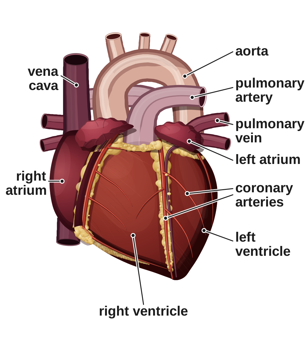
Screen 2
A real heart and a diagram
- A real heart is tilted, and you cannot see inside its chambers.
- A diagram is a simple, cut-open front view, so you can see all four chambers.
- In a diagram, red = oxygenated and blue = low in oxygen. Real blood is never blue.
Tap a difference to see it on both pictures.
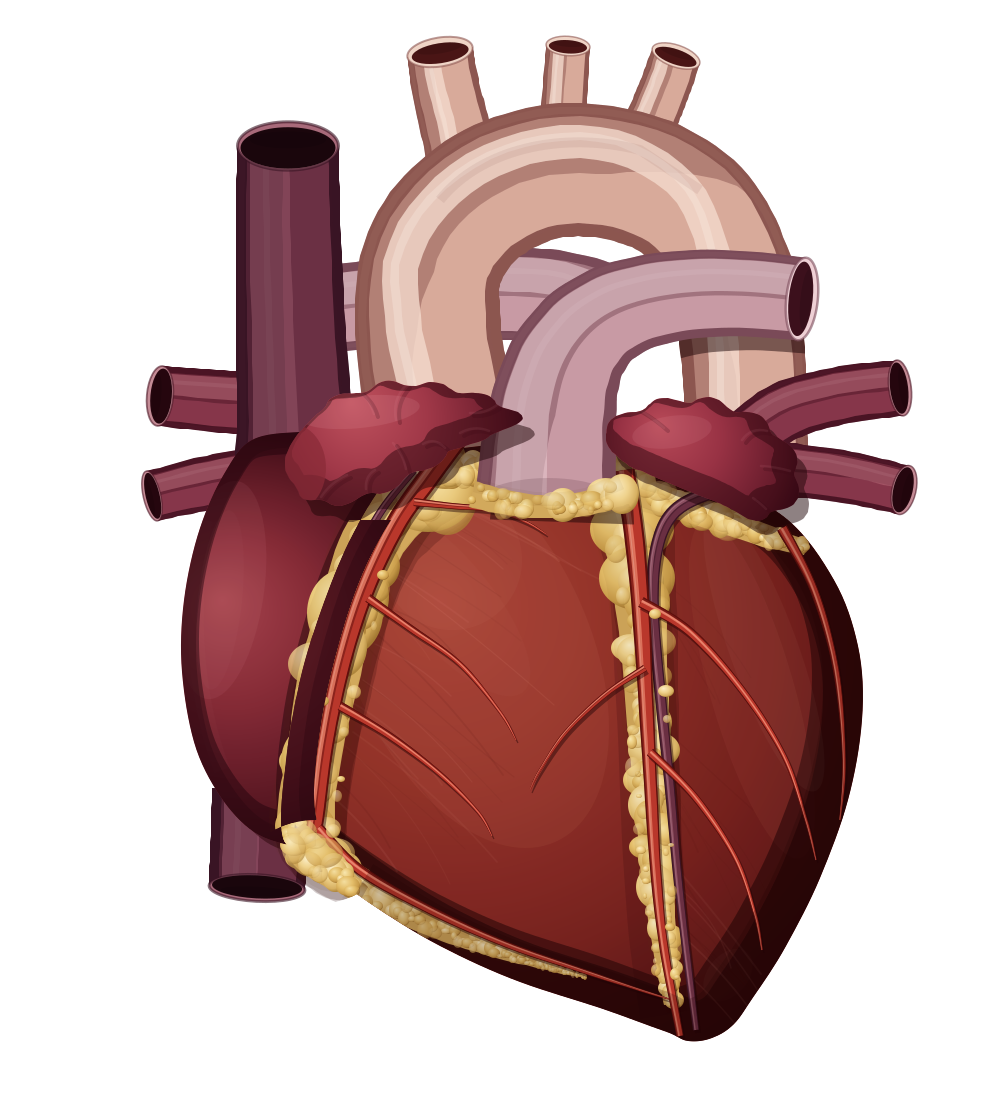
A real heart
right side ← | → left side
A diagram
right side ← | → left side
Screen 3
Inside the heart: four chambers
- Atria (top) receive blood. Ventricles (bottom) pump it out.
- Valves stop blood flowing backwards.
- Diagrams face you, so the heart's right side is on your left.
Tap a numbered pin to see its name for 3 seconds.
Screen 4
Watch the heart beat
- Blood flows into the atria. Pacemaker cells start each beat.
- The atria contract, then the ventricles contract and pump blood out.
- Valves shut so blood cannot flow backwards.
Screen 5
Checkpoint 1 of 3
Quick check on screens 1 to 4. Tap an answer.
1.In a heart diagram, some blood is coloured blue. What does the blue show?
2.In a heart diagram, where is the right atrium drawn?
3.What happens when the ventricles contract?
Screen 6
Blood vessels of the heart
- Arteries carry blood away from the heart. Veins carry blood back to the heart.
- In: vena cava (from the body), pulmonary vein (from the lungs). Out: pulmonary artery (to the lungs), aorta (to the body).
Arteries carry blood away from the heart. Veins carry blood back to it.
Screen 7
A double circulation
- Blood passes through the heart twice on each trip round the body.
- Right ventricle → lungs for gas exchange: blood picks up oxygen. Left ventricle → rest of the body.
Screen 8
Checkpoint 2 of 3
Quick check on screens 6 and 7. Tap an answer.
1.Which blood vessel carries blood from the heart to the lungs?
2.Where does the blood pick up oxygen?
3.How many times does blood pass through the heart on each trip round the body?
Screen 9
Two sides, two jobs
- The left ventricle has a thicker muscle wall. It pumps blood all round the body.
- More muscle → more force → higher pressure, so blood reaches the whole body.
- The right ventricle only pumps blood to the nearby lungs.
Screen 10
The heart's own blood supply
- Heart muscle contracts all the time. Its cells need oxygen for respiration, which releases energy.
- The coronary arteries branch off the aorta and supply the heart muscle with oxygenated blood.
Screen 11
Pacemakers and heart rate
- A group of pacemaker cells in the right atrium controls the natural resting heart rate.
- An artificial pacemaker is an electrical device that corrects an irregular heart rate.
- Heart rate (beats per minute) = number of beats ÷ time in seconds × 60.
Heart rate: 70 beats per minute (regular)
Work it out: Example: 21 beats in 15 seconds. 21 ÷ 15 × 60 = 84 beats per minute.
Screen 12
Checkpoint 3 of 3
Quick check on screens 9 to 11. Tap an answer.
1.Which chamber has the thickest muscle wall?
2.What do the coronary arteries do?
3.What is an artificial pacemaker?
Quiz
Quiz: B2 L6 The heart and circulation
10 questions. Tap an answer. Your first answer counts towards your score.
Which of these is true of a real heart, but NOT of a cut-open heart diagram?
In a diagram of the heart, where is the left ventricle drawn?
What is the job of the valves in the heart?
Which blood vessel brings oxygenated blood from the lungs back to the heart?
Which is the correct route for blood coming back from the body?
Why is the human circulation called a double circulation?
Why does the left ventricle have a thicker muscle wall than the right ventricle?
What do the coronary arteries do?
Where are the pacemaker cells that control the natural resting heart rate?
A student counts 20 heartbeats in 15 seconds. What is their heart rate?
Exam questions
The same five questions as the B2 L6 Exam questions worksheet. 20 marks.
Write your answer, then open the mark scheme and tick each point you made.
01Figure 1 shows a section through the human heart.
01.1Complete Figure 1.
Choose answers from the box.
01.2Which statement about the heart is correct?
Tick (✓) one box.
Mark scheme
- (1)
- (1)
- (1)
- (1)
01.1: 1 mark for each correct label. If more than one word from the box is written on one line, do not give that mark.
Model answer01.1 aorta; left atrium; right ventricle. 01.2 The right side of the heart pumps blood to the lungs.
02A student wanted to find their resting heart rate.
The student sat still for 5 minutes. Then they counted their heartbeats for 15 seconds.
They repeated this two more times.
Table 1 shows the results.
| Repeat | Number of heartbeats in 15 seconds |
|---|---|
| 1 | 16 |
| 2 | 18 |
| 3 | 17 |
02.1Calculate the mean number of heartbeats in 15 seconds.
02.2Calculate the student's resting heart rate in beats per minute.
Use your answer to Question 02.1.
02.3The natural resting heart rate is controlled by a group of cells.
Where are these cells found?
Tick (✓) one box.
02.4In some people these cells do not work properly, so their heart rate is irregular.
Name the electrical device that can be fitted to correct an irregular heart rate.
Mark scheme
- (1)
- (1)
- (1)
- (1)
- (1)
02.2: allow their 02.1 answer × 4, correctly calculated, for 2 marks. Correct answer with no working = 2 marks.
02.4: do not accept "pacemaker cells". do not accept "defibrillator".
Model answer02.1 (16 + 18 + 17) ÷ 3 = 17. 02.2 17 × 4 = 68 beats per minute. 02.3 In the right atrium. 02.4 An artificial pacemaker.
03Humans have a double circulatory system.
Describe what is meant by a double circulatory system.
Mark scheme
- (1)
- (1)
allow "blood goes from the heart to the lungs and back, then from the heart to the body and back" for 2 marks.
allow "one circuit from the heart to the lungs and one circuit from the heart to the body" for 2 marks.
allow "blood goes round two circuits / two loops" with no further detail for 1 mark.
do not accept "the heart has two sides" / "the heart beats twice" / "blood goes to the lungs twice".
Model answerBlood passes through the heart twice on each complete trip round the body: once on the way to the lungs and once on the way to the body.
04Figure 2 shows the outside of a heart.
The coronary arteries are on the surface of the heart.
A blood clot has blocked one coronary artery at point X.
The heart muscle in area Y stops contracting properly.
Explain why.
Mark scheme
- (1)
- (1)
- (1)
- (1)
max 3. allow "glucose" for oxygen. allow "no respiration". do not accept "energy is made / produced / created" (that point scores 0, others can still score). ignore "heart attack". ignore "the cells die".
Model answerThe clot stops blood flowing through the coronary artery to area Y. So the muscle cells in Y get little or no oxygen. They cannot carry out enough aerobic respiration, so not enough energy is released for contraction.
05The heart pumps blood around the body.
Explain how the structure of the heart is adapted for this function.
Mark scheme
Level of response. Read your answer, choose the level that fits best, then pick your mark.
| Level | Marks | Descriptor |
|---|---|---|
| 3 | 5–6 | A detailed and coherent explanation. At least three structures of the heart are each clearly linked to how they help it pump blood, with logical links, for example: thicker left ventricle wall → more force → higher pressure → blood reaches the whole body. |
| 2 | 3–4 | Several relevant structures are given and at least two are linked to their function. The links may be incomplete or not fully explained. |
| 1 | 1–2 | Simple statements about the structure of the heart, for example "it has four chambers" or "it is made of muscle", with little or no link to function. |
| 0 | 0 | No relevant content. |
Indicative content
- the heart is made of muscle, which contracts to pump blood
- the atria receive blood coming into the heart; the ventricles pump blood out of the heart
- the ventricles have thicker muscle walls than the atria, because they pump blood out of the heart (to the lungs or body) and the atria only pump blood into the ventricles
- the left ventricle has a thicker muscle wall than the right ventricle
- so it contracts with more force / produces a higher pressure, to pump blood all round the body
- the right ventricle has a thinner wall because it only pumps blood to the lungs, which are close by (and a lower pressure does not damage the lungs)
- valves prevent blood flowing backwards / make sure blood flows one way
- the wall between the two sides keeps oxygenated and deoxygenated blood apart, so the blood sent to the body carries as much oxygen as possible
- two sides / two pumps: the right side pumps blood to the lungs and the left side to the body (a double circulation)
- the veins (vena cava, pulmonary vein) bring blood into the atria; the arteries (pulmonary artery, aorta) take blood out of the ventricles
- coronary arteries supply the heart muscle with oxygen (and glucose) for respiration, to release energy for contraction
- pacemaker cells in the right atrium control the resting heart rate / start each heartbeat
Your mark:
Model answer (Level 3)The heart is made of muscle, which contracts to pump blood. The atria receive blood and the ventricles pump it out. The left ventricle has a thicker muscle wall than the right ventricle, so it contracts with more force and makes a higher pressure, which pushes blood all round the body. The right ventricle only pumps blood to the nearby lungs, so it needs less muscle. Valves stop blood flowing backwards, so blood flows one way through the heart. A wall down the middle keeps oxygenated and deoxygenated blood apart. The coronary arteries supply the heart muscle with oxygen for respiration, which releases the energy it needs to contract.
Total: 0 out of 20
Printable copy: the B2 L6 Exam questions worksheet and its mark scheme (PDF) are in .