Screen 1 of 12
Welcome back
Screen 1
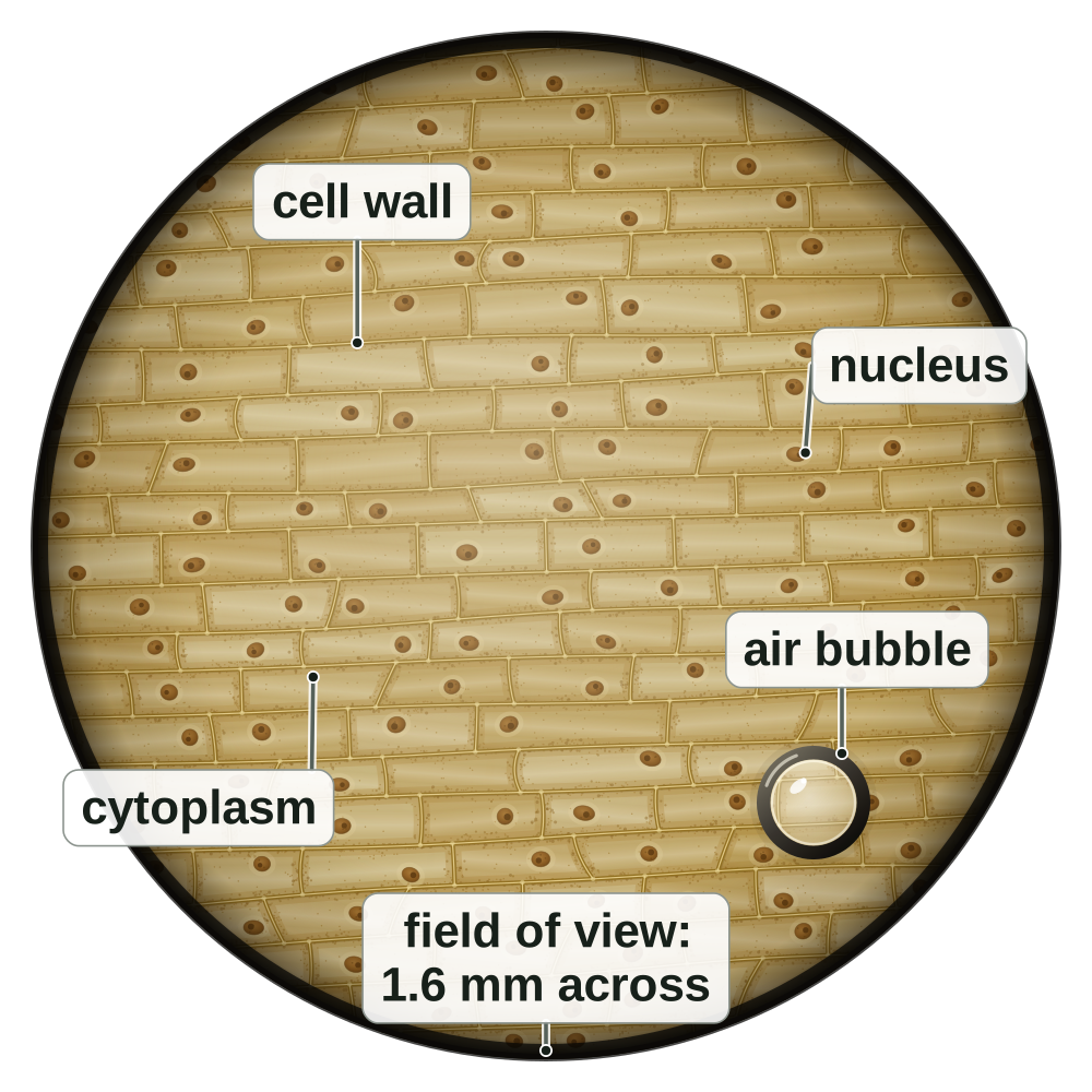
Onion cells at × 100
- This is a painting of the view down a light microscope at × 100.
- The cells are onion epidermis cells, stained yellow-brown with iodine solution.
- The circle is the field of view. At × 100 it is about 1.6 mm across.
- How big is one cell? This lesson shows you how to work it out.
Screen 2
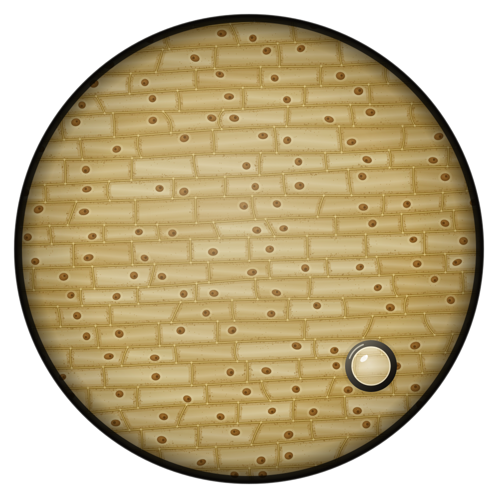
A real view and a drawing
- Left: the view down the microscope. Right: a biological drawing of the same cells, as in an exam.
- A drawing is simplified on purpose, and it has a scale bar.
- Exam pictures of cells are usually drawings like this.
Tap a difference to see it on both pictures.
The view at × 100
brick-shaped cells · walls · nuclei
A drawing
brick-shaped cells · walls · nuclei
Screen 3
Units for tiny things
- Cells are much smaller than 1 mm, so we use smaller units.
- 1 mm = 1000 µm (micrometres). 1 µm = 1000 nm (nanometres).
- To a smaller unit: multiply. To a bigger unit: divide.
Screen 4
How many times bigger?
- Magnification is how many times bigger the image is than the real object.
- magnification = size of image ÷ size of real object
- Magnification has no unit. Write it with a times sign: × 100.
Screen 5
Checkpoint 1 of 3
Quick check on screens 1 to 4. Tap an answer.
1.Down the microscope there is a round shape with a thick dark edge. Why is it left out of the biological drawing?
2.A human egg cell is about 0.1 mm across. How wide is it in micrometres (µm)? (1 mm = 1000 µm)
3.A drawing of a cell is 30 mm long. The real cell is 0.06 mm long. What is the magnification?
Screen 6
Finding the real size
- Cover what you want to find in the triangle. What is left tells you the sum.
- real size = image size ÷ magnification
- image size = real size × magnification
- Image size means the size of the image. Real size means the size of the real object.
Tap what you want to find.
Screen 7
Same units first
- Before you divide, put both sizes in the same unit. mm → µm: multiply by 1000.
- Try it on the calculator. Type the numbers only: never type the × sign into a calculator.
Screen 8
Checkpoint 2 of 3
Quick check on screens 6 and 7. Tap an answer.
1.Which equation gives the real size of an object?
2.A drawing of a cell is 18 mm long. The magnification is × 600. What is the real length of the cell?
3.A bacterium is 4 µm long. Its image in a micrograph is 12 mm long. What is the magnification?
Screen 9
Using a scale bar
- A scale bar is a line on a picture that shows a real length, such as 100 µm.
- Measure the bar and the cell with a ruler, in mm.
- real size = size on the picture ÷ length of the scale bar × value of the scale bar
Bar 20 mm, cell 40 mm. 40 ÷ 20 × 100 = 200 µm.
Screen 10
Cell size from the field of view
- At each magnification, the field of view has a known width (B1 L3 Using a microscope).
- Count the cells across the middle of the field of view.
- size of one cell ≈ width of the field of view ÷ number of cells across
| Magnification | Width of the field of view |
|---|---|
| × 40 | about 4.0 mm |
| × 100 | about 1.6 mm |
| × 400 | about 0.4 mm |
Screen 11
Standard form and orders of magnitude
- In standard form, a number is written as a number between 1 and 10, multiplied by a power of 10. A bacterium is 2 µm = 0.000 002 m = 2 × 10⁻⁶ m.
- Small numbers have negative powers of 10.
- Each order of magnitude is 10 times. An onion cell (200 µm) is 100 times longer than a bacterium (2 µm): 100 = 10², so 2 orders of magnitude.
Tap a thing to see its size in standard form, or compare two.
Screen 12
Checkpoint 3 of 3
Quick check on screens 9 to 11. Tap an answer.
1.On a drawing, a 50 µm scale bar is 10 mm long. A cell on the drawing is 30 mm long. What is the real length of the cell?
2.A field of view is 1.2 mm across. About 4 cells fit across it. About how long is one cell?
3.How is 0.000 005 m written in standard form?
Quiz
Quiz: B1 L4 Magnification and cell size
10 questions. Tap an answer. Your first answer counts towards your score.
Why are the onion cells in the painting yellow-brown?
Why does a biological drawing of cells have a scale bar?
A virus is 120 nm across. How wide is it in µm? (1 µm = 1000 nm)
What is the unit of magnification?
A student works out the real size of a cell. Their answer is bigger than the image. What have they most likely done?
An image of a chloroplast is 10 mm long. The magnification is × 2000. What is its real length?
A cell is 20 µm wide. It is drawn at a magnification of × 1500. How wide is the drawing?
On a micrograph, a 20 µm scale bar is 16 mm long. A nucleus is 8 mm wide. What is the real width of the nucleus?
At × 40, the field of view is 4 mm across. About 16 cells fit across it. About how long is one cell?
A grain of sand is 1 mm across. A bacterium is 1 µm long. How many orders of magnitude bigger is the grain of sand?
Exam questions
The same 5 questions as the B1 L4 Exam questions worksheet. 20 marks.
Write your answer, then open the mark scheme and tick each point you made. You may use a calculator and a ruler.
01This question is about the units used to measure cells.
01.1Draw one line from each unit to the length it is equal to.
Online: choose the length each unit is equal to from its list.
Unit
Equal to
- 10 mm
- 100 µm
- 1000 µm
- 1000 nm
01.2Which structure is the smallest?
Tick (✓) one box.
Mark scheme
- (1)
- (1)
- (1)
01.1: 3 correct lines = 2 marks; 1 or 2 correct lines = 1 mark (three correct: tick both boxes). If more than one line is drawn from a unit, none of its lines scores.
01.2: if more than one box is ticked, give 0 marks.
Model answer01.1 1 cm → 10 mm; 1 mm → 1000 µm; 1 µm → 1000 nm. 01.2 a ribosome, 25 nm wide.
02Figure 1 shows a student's drawing of a cheek cell.
The drawing was made at a magnification of × 400.
In Figure 1, the cell is 22 mm wide.
02.1Calculate the real width of the cheek cell.
Give your answer in micrometres (µm).
1 mm = 1000 µm
02.2What does "× 400" mean for the drawing in Figure 1?
Tick (✓) one box.
02.3Write your answer to Question 02.1 in metres.
Give your answer in standard form.
Type the number in the first box and the power (for example −4) in the small box.
Mark scheme
- (1)
- (1)
- (1)
- (1)
- (1)
02.1: correct answer with no working = 3 marks. allow 22 000 µm ÷ 400 = 55 µm (units changed first) for 3 marks. 0.055 mm = 2 marks; 0.055 with the unit µm = 2 marks (the unit was not changed). do not accept 8800 (22 × 400: multiplied).
02.2: if more than one box is ticked, give 0 marks.
02.3: allow their 02.1 answer correctly written in metres in standard form. do not accept 55 × 10⁻⁶ m or 0.000 055 m (not standard form). do not accept 5.5 × 10⁵ m.
Model answer02.1 22 ÷ 400 = 0.055 mm; 0.055 × 1000 = 55 µm. 02.2 The drawing is 400 times bigger than the real cell. 02.3 55 µm = 0.000 055 m = 5.5 × 10⁻⁵ m.
03Figure 2 shows a drawing of a bacterium with a scale bar.
In Figure 2, the scale bar is 30 mm long and the bacterium is 45 mm long.
Calculate the real length of the bacterium.
Mark scheme
- (1)
- (1)
correct answer with no working = 2 marks. allow 45 ÷ 15 = 3 µm (15 mm stands for 1 µm). do not accept 2 µm (the scale bar's label). ignore units.
Model answer45 ÷ 30 = 1.5; 1.5 × 2 µm = 3 µm.
04A student uses a light microscope to look at the cells in a moss leaf.
On this microscope, at a magnification of × 100, the field of view is 1.8 mm across.
The student counts 30 cells across the middle of the field of view.
04.1Estimate the length of one moss leaf cell.
Give your answer in µm.
04.2The student changes the magnification to × 400.
Explain why the student sees fewer cells across the field of view.
Mark scheme
- (1)
- (1)
- (1)
- (1)
04.1: correct answer with no working = 2 marks. allow 1800 µm ÷ 30 = 60 µm. 0.06 mm = 1 mark. do not accept 54 (1.8 × 30).
04.2: allow "the field of view is only about a quarter as wide (about 0.45 mm)" for the first mark. allow "only about 7 or 8 cells fit across" for the second mark. do not accept "the cells get bigger" (the real cells do not change; their image does).
Model answer04.1 1.8 ÷ 30 = 0.06 mm = 60 µm. 04.2 At × 400 the field of view is smaller: only about a quarter as wide, about 0.45 mm across. Each cell looks bigger and takes up more of the field of view, so fewer cells fit across it.
05A student has two micrographs.
- Micrograph A shows plant cells at a magnification of × 200.
- Micrograph B shows bacteria at a magnification of × 20 000.
The student wants to find out how many times bigger a plant cell is than a bacterium.
Describe how the student could use the two micrographs to find this out.
Mark scheme
Level of response. Read your answer, choose the level that fits best, then pick your mark.
| Level | Marks | Descriptor |
|---|---|---|
| 3 | 5–6 | A clear, logical method. Both cells are measured with a ruler, both real sizes are calculated with real size = image size ÷ magnification (or magnification = size of image ÷ size of real object, rearranged), both are put in the same unit, and the plant cell's size is divided by the bacterium's size. The steps are in a logical order. (A way to improve the result or to check it is sensible is credit-worthy but not needed for Level 3.) |
| 2 | 3–4 | Measuring and using the magnification for at least one micrograph, with an idea of comparing the two sizes. One step, such as changing to the same unit, may be missing or unclear. |
| 1 | 1–2 | Simple statements, such as "measure the cells" or "the plant cell is bigger", with no clear method. |
| 0 | 0 | No relevant content. |
Indicative content
- measure the length of a plant cell in micrograph A with a ruler (in mm)
- measure the length of a bacterium in micrograph B with a ruler (in mm)
- real size = image size ÷ magnification
- plant cell: divide its image length by 200
- bacterium: divide its image length by 20 000
- change both real sizes into the same unit (such as µm: × 1000)
- divide the real size of the plant cell by the real size of the bacterium
- the answer is how many times bigger the plant cell is
- measure several cells of each and use the mean
- measure the same dimension each time (such as the longest length)
- give the answer as an order of magnitude (for example, about 100 times = 2 orders of magnitude)
- check the answer is sensible: a plant cell is about 10 to 100 µm, a bacterium about 1 to 5 µm
Your mark:
Model answer (Level 3)Use a ruler to measure the length of one plant cell in micrograph A, in mm. Work out its real length: real size = image size ÷ magnification, so divide the image length by 200. Do the same for one bacterium in micrograph B, dividing its image length by 20 000. Change both real lengths into µm by multiplying by 1000, so they are in the same unit. Then divide the real length of the plant cell by the real length of the bacterium. This tells you how many times bigger the plant cell is. For example, a plant cell image 20 mm long gives 20 ÷ 200 = 0.1 mm = 100 µm, and a bacterium image 40 mm long gives 40 ÷ 20 000 = 0.002 mm = 2 µm, so the plant cell is 100 ÷ 2 = 50 times longer. To make the answer more reliable, measure several cells of each type and use the mean. Check it is sensible: a plant cell should be about one to two orders of magnitude bigger than a bacterium.
Total: 0 out of 20
Printable copy: the B1 L4 Exam questions worksheet and its mark scheme (PDF) are in Downloads.
Downloads
- PPTXB1 L4 PowerPointLesson with checkpoints, revision notes, all worksheets and answers in green. 7.0 MB.
- PDFB1 L4 Revision notes4 printable pages, 0.7 MB.
- PDFB1 L4 Mandatory worksheetThe main worksheet (Foundation). 2 pages, 0.3 MB.
- PDFB1 L4 Challenge worksheetHarder questions. 2 pages, 0.2 MB.
- PDFB1 L4 Alternative worksheetKey notes and simpler tasks. 2 pages, 0.4 MB.
- PDFB1 L4 Exam questionsExam-style, black and white, 5 questions, 20 marks: the same questions as the online exam questions. 4 pages, 83 KB.
- PDFB1 L4 Exam questions mark schemeThe mark scheme for the exam questions. 2 pages, 47 KB.
Worksheet answers are in the PowerPoint, in green, after each worksheet.